Posts

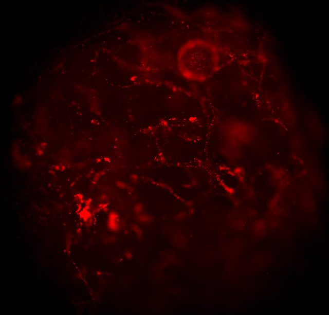
Extracellular Ca2+-sensing fluorescent protein biosensor based on a collagen-binding domain

Our group published new research article, focused on further development of the 'biosensor scaffold' concept for advanced tissue engineering applications, such as imaging-assisted organoid engineering. Here, we collaborated with colleagues from Agilent Technologies and University of Tubingen (Laboratory of Professor Katja Schenke-Layland, Germany) to develop and functionally evaluate recombinant extracellular Ca2+-specific protein biosensors that can bind collagen- and cellulose-based tissue engineering scaffold materials. Using confocal and two-photon FLIM, and Matrigel-based 3D culture of mouse intestinal organoids, we evaluated performance of the collagen-binding biosensor and found that 3D culture of the intestinal epithelium can regulate extracellular Ca2+, potentially through the function of lipid droplets. The article published in ACS Applied Bio Materials journal can be assessed here . Twitter link . 

Disruption of hypoxia-inducible fatty acid binding protein 7 induces beige fat-like differentiation and thermogenesis in breast cancer cells

Dr. Ruslan Dmitriev  has co-authored a paper focused on molecular mechanisms of breast cancer and the role of mitochondrial function in thermogenesis. This work, which is a result of collaborative efforts  from Oxford University, Kyoto University, University College Cork and Sechenov Medical University research groups, has been recently published in Cancer & Metabolism journal. Full-text is available here . Twitter link .

Giants in sensing (accordingly to ACS Sensors journal)

Image
Our recent collaborative work on pH-sensing DAOTA dyes (published in Analytical Chemistry in 2019) has been included into the virtual issue of ACS Sensors journal, celebrating 5 years and entitled 'Giants in sensing'. To reflect back to what we have published over the period of 2015-2020 with ACS journals, see the list below: Dmitriev RI , Borisov SM, Düssmann H, Sun S, Müller BJ, Prehn J, Baklaushev VP, Klimant I, Papkovsky DB: Versatile Conjugated Polymer Nanoparticles for High-Resolution O2 Imaging in Cells and 3D Tissue Models.  ACS Nano  2015,  9 (5), 5275-5288. Dmitriev RI , O’Donnell N, Papkovsky DB:   Metallochelate Coupling of Phosphorescent Pt-Porphyrins to Peptides, Proteins, and Self-Assembling Protein Nanoparticles .   Bioconjugate Chemistry   2016,   27 (2):439-445. Fomin M,  Dmitriev RI , Jenkins J, Papkovsky DB, Heindl D, König B: A Two-Acceptor Cyanine-Based Fluorescent Indicator ...

Updates during COVID-19 pandemic

Image
We are still alive and continue to work! Over the past month of nation-wide work restrictions and subsequent lockdown our team did not stay idle and we participated in a number of research, collaborative and teaching activities. - Shortly before the lockdown we finalised our new manuscript reporting new biosensor scaffold materials reporting extracellular calcium dynamics in 3D organoid model. The preprint version is available on BioRxiv . - Together with Dr. M. Monaghan group (Trinity College Dublin) Dr. Dmitriev co-authored a review book chapter on optical imaging approaches employed in tissue engineering. This book chapter has been published as part of Springer Reference Series in Biomedical Engineering . The text (free to the members of TERMIS) is available here . Twitter link - Members of the team in collaboration with Hubrecht Institute has prepared a book chapter for a book 'Intestinal Stem Cells', which is edited by Paloma Ordonez-Moran and will b...

New publication from the group: imaging of O2 nanosensors in implanted scaffold materials

Image
Dr. R. Dmitriev  performed and co-authored a collaborative study together with groups from Russia (Sechenov Institute for Regenerative Medicine and Privolzhsky Research Medical University) and Austria (TU Graz), which describes multi-parameter imaging of decellularized pericardial biomeshes in an implanted mouse model. One of highlights of the study is the finding that conjugated polymer-based O2 nanosensors can bind and remain bound to the biomeshes in live animal for extended period of time and thus used for subsequent O2 imaging in live animal and ex vivo. This confirms high usability of conjugated nanoparticle sensors previously developed by our team for such emerging applications as monitoring of tissue regeneration, tumour imaging and others. The work has been published in Frontiers in Bioengineering and Biotechnology journal. Full text can be assessed here . Twitter link .

Biophysical Society 2020, San Diego

Image
Dr. Ruslan Dmitriev  recently presented a talk at the Biophysical Society 2020 Annual Meeting, in San Diego Convention Center, California, USA. In his talk, Ruslan summarised recent group developments in a multiparameter imaging of stem cell metabolism in the small intestinal organoid model. He focused on measuring oxygenation of live intestinal organoids, assessing mitochondrial polarisation and combining O2 with NAD(P)H redox microscopy analyses. He also received  Travel Award from the Biophysical Society to attend this meeting. More information on the meeting can be found on Twitter .  The abstract of presentation published in Biophysical Journal supplement can be viewed here .

Interview to Biotechniques journal

During the ASCB-EMBO 2019 conference in Washington DC (December 2019) Dr. Ruslan Dmitriev was interviewed by Biotechniques journal about FLIM, organoids, challenges and career inspirations. Here's an interview: https://www.biotechniques.com/microscopy-imaging/ruslan-dmitriev-on-flim-and-intestinal-organoids/