Posts

TERMIS-European conference 2022 in Krakow

Image
 We are proud to organise a symposium at TERMIS-EU 2022 conference in Krakow, Poland! More information on TERMIS can be found here: https://eu2022.termis.org/call-for-abstracts/

Database on multicellular spheroids

Image
Our team contributed to the database summarising features of multicellular spheroids, produced from cancer and non-transformed cells.  3D spheroid cell cultures often suffer from incomplete reporting, lab-to-lab variability and other factors, which can affect reproducibility of research. The multidisciplinary team led by Prof. Olivier De Wever (LECR, CRIG) performed analysis of factors affecting spheroid reproducibility - among the others, spheroid formation method and growth medium composition and glucose content were found to be among the most important. The work published in Nature Methods journal can be assessed here . The  News and Views  feature. Feel free to use the database ! https://www.mispheroid.org 

TERMIS-World Congress 2021

Image
  We are not presenting this year and the TERMIS world congress is virtual but we are still participating! - Prof. Dmitriev will co-chair the session ET2.2. on ' Advanced spatiotemporal imaging in regenerative medicine' (November, 17th, 15.30-17.00). In this session, Prof. Michael Monaghan (Trinity College Dublin) is also going to talk about our recently published joint research on microfluidic-based perivascular stem cell niche modelling . Stay tuned!

FLIM and PLIM across three dimensions in biological applications (review)

Image
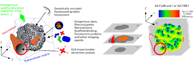
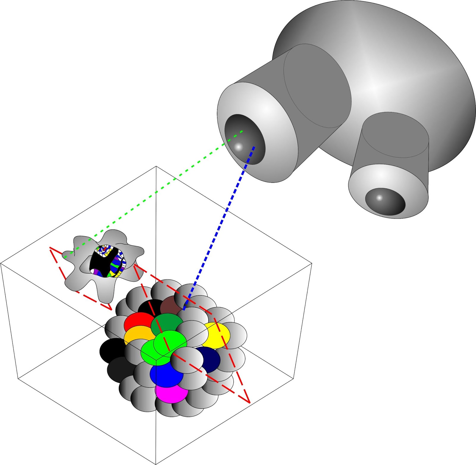
Together with Prof. Margarida Barroso (Albany Medical College, NY) and Prof. Xavier Intes (Rensselaer Polytechnic Institute, NY) we wrote a review article summarising biological aspects of applications of the fluorescence (FLIM) and phosphorescence (PLIM) lifetime imaging microscopies.  We covered most of FLIM and PLIM applications, including FLIM-FRET, macro-FLI, O2 and metabolic imaging, monitoring virus entry into the cell, imaging in organoids and tissue engineering and other areas. In addition, we outlined broad applicability of this type of microscopies not only for cancer and stem research (biomedicine) but also for fungal, plant and other 'general biology' areas.  The review published in the Journal of Cell Science can be assessed here . Twitter link  and link2

JoVE Methods collection: Quantitative live cell imaging of 3D models

Image
Together with Prof. Margarida Barroso (Albany Medical College), we guest edit a collection of video protocols, hosted by the Journal of Visualised Experiments ( JoVE ), and  dedicated to the quantitative live cell imaging in 3D .  We expect bringing together some leaders and innovators in various aspects of the quantitative live fluorescence microscopies applied to ex vivo animal models, organoids and 3D engineered tissue blocks. The collection, which just has been started, can be assessed here .

Intracellular label-free detection of mesenchymal stem cell metabolism within a perivascular niche-on-a-chip

Our team has co-authored an article focused on studying oxygenation and cell metabolism in bioreactor mimicking perivascular stem cell niche ('niche-on-a-chip'). This study combines mathematical modelling of flow and oxygenation of mesenchymal stem cells with actual analysis of their metabolism via multiparameter analysis of NAD(P)H (two-photon FLIM), cell oxygenation (two-photon ratiometric fluorescence), mitochondrial polarisation and glucose transport. The study is a result of joint efforts of Politecnico di Milano, Trinity College Dublin, NUI Galway and Ghent Univeristy. The article published in Lab on a Chip journal can can be assessed here . Twitter link

Second Human Structure and Repair research symposium

Image
Prof. R. Dmitriev will be presenting at HSR research symposium (Ghent University, 11-Feb-2021) and speak about new biosensors for organoid research. Full details and registration can be found here:  https://www.ugent.be/ge/hsr/en/news-events/events/2nd-hsr-research-symposium-2021