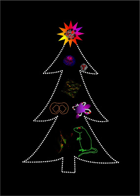
Crown ethers help to design advanced nanobiosensors for 3D tissue models
Fluorescence imaging helps studying
biological processes in live cells, tissues, animals and humans in a minimally
invasive way, to advance the basic, cancer and brain research, from the lab to
the bedside. State-of-the-art biosensing nanoparticles are renown for
exceptional brightness and stability, unmatched by the fluorescent dyes and
fluorescent proteins. However, even such advanced materials as nanoparticles
often suffer from poor performance, complex interaction with biomolecules and
cells, when challenged with real biological models, such as brain tissue.
In search of new ways of improving brain
tissue staining with polymer nanoparticles, the collaborative team including
researchers from University College Cork, Graz University of Technology
(Austria), University of Maryland (USA) and Empa (Switzerland) and led by Dr. Ruslan Dmitriev discovered that widely known RL100 cationic polymer can be
successfully combined with the new generation of deep red fluorescent
ion-selective biosensors called fluoroionophores. This innovative fluorescent nanomaterial,
combining hydrophobic and charged moieties, has strong ability to stain the
broad range of cells, including ‘difficult-to-grow’ neurons and glial cells.
For the first time, the efficient staining of 3D tissue models was shown, which
included tumor- and stem cell-based spheroids, organoids and brain sections.
After thorough study, confirming the mechanism of their internalization and
intracellular stability, the Team applied fluoroionophore nanoparticles for
imaging of potassium-induced changes in membrane potential in neuronal cell
cultures, and in vivo epileptic seizures in the brain. Developed nanoparticles
are highly useful tool as advanced biosensor for imaging of potassium ion
dynamics in neuronal cells and the living brain, bioenergetics and drug
screening. They can be used as a platform for advanced design of future polymer-based
nanoparticle biosensors.
The research has been published in Advanced
Functional Materials journal on 5-Jan-2018. The full text can be assessed here.
More on this story in the news can be found here.


Comments
Post a Comment