Posts

Strategic workshop on imaging subcellular to cellular cancer biology

Image
Dr. R. Dmitriev participated in Strategic workshop on cancer biology (subcellular to cellular cancer imaging) held at National Institutes of Health, Bethesda MD, USA on 5-6 April 2018. The event helped to network some leaders in quantitative imaging of cancer across the scales, from cell cultures to advanced in vivo imaging, consolidating the knowledge in two-photon intravital, imaging 3D tissue models, biosensors, FLIM, label free methodologies and others. The event was organised by National Cancer Institute, USA and the American Society for Cell Biology (ASCB). Dr. R. Dmitriev presented a poster summarising recent developments of FLIM and PLIM for multi-parametric live imaging of 3D tissue models: The event webpage Twitter link .

Europt(r)ode XIV, Napoli 2018

Image
Members of the lab will attend the Europt(R)ode conference on 25-28 Mar 2018. They will discuss multi-parametric FLIM-PLIM imaging of 3D tissue models (Dr. R. Dmitriev, regular talk) and biomedical applications of phosphorescent oxygen sensors and probes (Prof. D. Papkovsky, invited talk). Europt(R)ode is one of the leading worldwide conferences focusing on innovations in optical sensors and biosensors and their multi-disciplinary applications. More information on the conference and the program can be found here .

Stop codon readthrough generates a new vitamin D receptor variant

Dr. R. Dmitriev collaborated with Dr. G. Loughran, Prof. J. Atkins (School of Biochemistry and Cell Biology, UCC) and other colleagues from National Institutes of Health (USA), University of Tubingen (Germany), National Institute for Bioprocessing Research and Training (Ireland) and MIT (USA), to understand the phenomenon of stop codon readthrough in vertebrates. The team found that human mRNA with stop codon motif UGA_CUAG displayed inefficient termination of translation of vitamin D receptor (VDR) in 6.7% cases, leading to translation of extended polypeptide product, in so-called 'readthrough' phenomenon. Interestingly, this longer variant of VDR, termed as VDRx also was functional but had reduced activity. The importance of this discovery is still to be understood. The results of study published in the Journal of Biological Chemistry can be found here .

Twitter

Image
Metabolic Imaging Group & Laboratory of Biophysics and Bioanalysis can be found on Twitter now! Make sure to follow our channel  @ PhotoBioLab  to stay informed about our latest accomplishments!

Crown ethers help to design advanced nanobiosensors for 3D tissue models

Image
Fluorescence imaging helps studying biological processes in live cells, tissues, animals and humans in a minimally invasive way, to advance the basic, cancer and brain research, from the lab to the bedside. State-of-the-art biosensing nanoparticles are renown for exceptional brightness and stability, unmatched by the fluorescent dyes and fluorescent proteins. However, even such advanced materials as nanoparticles often suffer from poor performance, complex interaction with biomolecules and cells, when challenged with real biological models, such as brain tissue. In search of new ways of improving brain tissue staining with polymer nanoparticles, the collaborative team including researchers from University College Cork, Graz University of Technology (Austria), University of Maryland (USA) and Empa (Switzerland) and led by Dr. Ruslan Dmitriev discovered that widely known RL100 cationic polymer can be successfully combined with the new generation of deep red fluorescent ion-se...

Lab members in the news

Prof. Dmitri Papkovsky and Dr. Ruslan Dmitriev have recently appeared in the news on a website of the School of Biochemistry/ University College Cork. Prof. Papkovsky's two-weeks lecture trip across Japanese universities, supported by the Japan Society for Promotion of Science has been mentioned here . Dr. Dmitriev has appeared in two news articles, mentioning his new book 'Multi-parametric live cell microscopy of 3D tissue models' published by Springer, and about his new role as ASCB ambassador .

Multi-Parametric Live Cell Microscopy of 3D Tissue Models (book)

Image
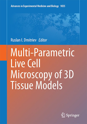
Dr. Ruslan Dmitriev edited the book 'Multi-parametric live cell microscopy of 3D tissue models ', recently published by Springer publishing house (eBook available from 30-Oct-2017, printed version available from Nov-2017), as part of renown  Advances on Experimental Medicine and Biology series (vol. 1035).  The book brings together some world-known experts and pioneers in the area of multi-parametric live cell microscopy and imaging of 3D tissue models in tissue engineering, neurobiology, stem cell and cancer biology, to provide and introduction and easy-to-follow experimental methods. The instrumentation and data analysis, scaffold and types of 3D tissue models, choice of biosensor design as well as examples of applications and methods are included in this book. Members of Metabolic Imaging Group and Biophysics and Bioanalysis Lab contributed in three chapters covering: -Overview of 3D models and available biosensors (Chapter 4): https://link.springer.com/chap...