Posts

Publication alert: Phasor-FLIM visualizes and quantifies polluting nanoplastic particles in live intestinal epithelium

Image
  Increased micro- and nanoplastic (MNP) pollution and its poorly understood biomedical impact raises questions on their cell and tissue uptake mechanisms and effects on the absorptive tissues. Dmitriev’s team reports a new microscopy-based approach to perform dynamic imaging of dye-modified micro- and nanoplastics (MNP). When combined with stem cell-derived intestinal organoids, with controlled apical and basal polarity, this approach enables for sensitive detection and quantification of MNPs, their internalization and biological effects.  The work was published in Light: Science & Applications , big thanks to all collaborators and researchers involved, especially Irina, Hang (first 'big paper' in PhD) and Bert! We hope to get this research line funded and expand towards studying effects of MNPs on the brain, tooth development and other tissues! Press-release on Eurekalert (14-09-2025)  

Lab Photo: June 2025

Image
Up to date photo of our team!   Nore, Irina, Johanna, Magali, Angela, Michele, Gabriele, Teresa*, Sara* and Hang First row: Ruslan  *Sara and Teresa are visiting PhD students funded by MSCA-DN Horizon project FLIMagin3D. 

New publication: evaluation of GelMA as a Matrigel replacement for small intestinal organoids

Image
      In our new study (collaboration between Ghent University and Rousselot BV) we report application of GelMA (gelatin methacryloyl) hydrogels with tuneable mechanical properties and added laminin to the culture of the small intestinal organoids. We found that mechanical properties alone are not sufficient for basement membrane support, resulting in polarity reversion, in contrast to the Matrigel-grown cultures. Altogether, observed polarity reversion phenomenon highlights importance of the further research in biochemical factors, directing cell polarity in the epithelial cells, while it also provides a tractable way for producing 'apical-out' organoids. Congratulations to Lenie (for her work on this MSc research project), Irina (for her supervision and driving force), Thomas and Ruben for expertise with gelatins and Bert for expertise with pig intestinal organoids. The work has been published in the Journal of Tissue Engineering (Open Access): https://journals.sagepub...

New synthetic hydrogels for tumour spheroids and intestinal organoid cultures (publication)

Image
  We collaborated with groups of Prof. Richard Hoogenboom and Prof. Devriendt (Ghent University) and tested application of poly(2-alkyl-2-oxazoline) hydrogels (PAOx) for 3D cell cultures of cancer and stem cells. We found that encapsulation of small intestinal organoids into PAOx hydrogels results in polarity reversion, i.e. transition from basal to apical-out phenotype. This is useful for studying drug delivery, nutrient absorption and host-microbiota interactions within this model.  This figure shows dramatic differences of mitochondrial polarisation (TMRM) and appearance (transmission light) in small intestinal organoids, observed after apical-out transition (PAOx), in contrast to conventional Matrigel-grown culture: Since the PAOx hydrogels can show variable mechanical properties, we tested if encapsulation into stiff and soft hydrogels can affect behaviour of cancer cells in 3D: indeed, with HCT116 spheroids we found that stiffness of the hydrogels affect cell migration f...

Happy New Year 2025! New publications.

Image
Wow, time really does fly! It's been a while since our previous update but by no means our team was idle.  End of 2024 was memorable with exciting conferences, including SPAOM 2024, Histochemical Society Annual online meeting 2024, FHERITALE consortium and others! We've received funding to study neurodevelopmental and neurodegenerative diseases (more news soon) so we can plan new & exciting research projects. Our team currently hosts 2 international researchers, Valerie Cote and Dr. Simone Perottoni, both learning and doing exciting science in FLIM, organoids and imaging of cancer cells / tissues. In addition, we are looking forward to host more of PhD students from FLIMagin3D consortium! Here are some recent publications, from our team and collaborative research projects: Sheehan A, Okkelman IA, Groslambert G, Bucher C, Dmitriev RI, Filatov MA: Optoelectronic Properties and Fluorescence Lifetime Imaging Application of Donor-Acceptor Dyads Derived From 2,6-DicarboxyBODIPY ...

See the hidden: discover more with FLIM (online event)

Image
Prof. R. Dmitriev will participate in online event 'See the hidden: discover more with FLIM' on 10-Oct-2024! More info & registration can be found here: https://events.bitesizebio.com/see-the-hidden-discover-more-with/join

Conference presentations: September 2024

Image
Our team is growing and we can present at the same time in two different places now! PhD candidate Nore Van Loon (joint project with FunGen lab, Prof. S. Vergult) gave a talk at the Methods and Applications of Fluorescence 2024 conference (8-11 September Valencia, Spain)! She presented her work in progress on using autofluorescence and fluorescent cholera toxin labelling to monitor differentiation of neural cells in human iPSC-derived 'brain' organoids. This work is a part of ongoing efforts of bringing together multi-omics approaches and advanced imaging of organoids to better understand aetiology of such neurodevelopmental disorders as FOXG1 syndrome, funded by the Special Research Fund at Ghent University (BOF) and Marguerite-Marie Delacroix foundation. At the same time, Prof. Ruslan Dmitriev travelled to Munster, Germany, to present an invited lecture on imaging oxygenation and cell metabolism in multicellular spheroids in 4th Inflammation & Imaging symposium! This l...

Production and Multi-Parameter Live Cell Fluorescence Lifetime Imaging Microscopy (FLIM) of Multicellular Spheroids (JOVE protocol)

Image
ALERT: New publication from our group! Angela, Gabriele, Hang, Nore, Nina and Irina teamed up to produce a video protocol paper, focusing on comparing various multicellular spheroid formation methods, suitable for 'downstream' live multi-parameter fluorescence lifetime imaging microscopy (FLIM) and imaging cell oxygenation. This protocol is illustrated with examples of autofluorescence-based FLIM microscopy of NAD(P)H, oxygenation and cell death, as well as imaging of visible flavin-based cell autofluorescence, performed in tumour and stem cell-derived spheroids. The article is also a part of the Methods collection 'Quantitative Live Cell Imaging of 3D Models', which is going to expand in the future.    Link to the publication: https://www.jove.com/t/66845/production-multi-parameter-live-cell-fluorescence-lifetime-imaging The Methods Collection (Journal of Visualised Experiments, JOVE):  https://app.jove.com/methods-collections/902

End of the Academic Year 2023-2024!

Image
It's been busy, it's been tough but it's been also fun! Our team has trained a number of MSc students, with excellent projects, spanning the scope from establishment of novel organoid models, to testing of mechanical properties of the hydrogel-producing biomaterials on the small intestinal epithelium organoids and FLIM analysis of neural ('brain') iPSC-derived organoids. Thanks to everyone involved! The end of year (June) was culminated in presenting of research at Ghent Gut Inflammation Group (Ruslan, Irina, Hang, Lenie) and participating (as instructors, Ruslan and Michele) in the FEBS course on Functional Imaging of cellular dynamics (Amsterdam, 9-15 June 2024)!         Stay tuned for more exciting news on our research!

Brief updates for Academic Year of 2023-2024

(video: time lapse FLIM of endocytosis in human colon cancer cells using LysoSensor Green, (C) Dmitriev Lab, 2023) The period of ~October 2023 until ~May 2024 (almost complete academic year!) was too busy for publishing any updates but thanks for staying tuned (if you did so). Some key updates for this period: Successful start of FLIMagin3D project, with the 3 new PhD students (Hang, Michele and Gabriele) + visiting (via secondments) PhD students, joining our team to address challenges in multi-parameter FLIM of organoids and multicellular spheroids, advancing imaging & probes for cell oxygenation, FLIM-FRET, tissue mechanics and biofabricated inter-species communities. More about the FLIMagin3D can be found here:  https://www.flimagin3d.com/   Some visiting scientists with exciting projects, including Dr. A. Russegger, R. Harkhoe, partners from OsteoNet Horizon MSCA Staff Exchange network Our group (including the microscopes!) has relocated to the new 'The Core' / MREB3 r...

TERMIS-EU 2023

Image
We will attend and present at TERMIS-EU 2023 (Manchester, UK, 28-31 March 2023)! Prof. R. Dmitriev will co-chair (together with Prof. M. Monaghan, Trinity College Dublin) and present at the symposium ' Wanted: dead or alive? Quantitative microscopy of single-cell, spheroid and organoid cultures': Fluorescence Lifetime Imaging Microscopy (FLIM) to study niche-specific regions and metabolism in epithelial organoids More on the program and the event can be found here .

Research on nanothermometry continues (new paper from the team)

Prof. Ruslan Dmitriev and PhD candidate Angela Debruyne collaborated with TU Graz (Prof. S. Borisov) and University of Tubingen on the design of new type of the nanothermometer, based on organic Zr complexes and exhibiting long emission decay times. These materials open a new framework for microsecond-range emitting temperature-sensitive materials, providing better resolution and potential applications in imaging temperature gradients in multicellular spheroids, as we have demonstrated in the study. The research has been published in Advanced Optical Materials journal. Russegger A, Debruyne AC, Berrio DC, Fuchs S, Marzi J, Schenke-Layland K, Dmitriev RI, Borisov SM: Bright and Photostable TADF-Emitting Zirconium(IV) Pyridinedipyrrolide Complexes: Efficient Dyes for Decay Time-Based Temperature Sensing and Imaging . Advanced Optical Materials 2023,:2202720. If you are interested in our research in nanothermometry, expertise and dedicated nanosensors, do not hesitate to contact us. ...

FLIMagin3D network (we are hiring!)

Image
Are you looking for a PhD and interested in advanced live cell microscopies? Perhaps in FLIM? In some of the best labs across EU and UK? With funded mobility? We have good news for you: within the Horizon-funded MSCA doctoral network we are hiring 12+2 PhD students. More information here . Apply via EURAXESS / FLIMagin3D website or for UK here  Twitter 

Updates from the group (October 2022)

Image
It was a busy period during June-October this year! Below are some important updates about our team: - Together with prof. Sarah Vergult, we received a funding by the Special Research Fund (BOF) to start an interdisciplinary research project on imaging of neural organoids 'IMAGIBRAINDD' . The team will grow with at least one new PhD student. More details to follow. - Have you ever heard about FLIMagin 3 D ? Together with Prof. Michael Monaghan (Trinity College Dublin) and a number of experts across EU and UK we have secured funding for a new doctoral training network (MSCA) aiming at expanding & consolidating training in biosensors, modelling, tissue models, hardware and software for 3D FLIM. This project will start from 1-Jan-2023 and soon we will be hiring PhD students. Twitter page for new updates. - New microscope(!) Thanks to FWO (medium infrastructure call 2021) and support from ~ 25 other enthusiastic colleagues from UGent and VIB, soon we will have confocal white ...

Lab photo, May 2022

Image
Tissue Engineering and Biomaterials Research Group @UGent in May 2022:  From left to right: W. Vandenberghe, M. Meul, J. Aernoudt, R. Dmitriev, I. Okkelman and A. Debruyne

Affordable microscopy of spheroid oxygenation (video protocol)

Image
Our lab has published a new protocol paper, this time with the video, in the Journal of Visualized Experiments (JoVE)! In this research method paper, we report the use of red/ near-infrared emitting biocompatible nanosensors to monitor oxygenation of the live multicellular spheroids (made of one or more cell types), together with the tracers of cell death. While this methodology is typically available only for users with 'high-end' PLIM microscopes and alike, we show that using of ratiometric readout with the red and near infrared emitting fluorescent nanosensors makes this possible on broadly available 'widefield' fluorescence microscope platforms. This method helps comparing oxygenation of spheroids, assessing this in a long-term and kinetic assays, comparing their variability and viability, essential in follow-up 3D printing applications. This is the first article in our collection ' Quantitative live cell imaging of 3D models' , guest-edited together with ...

TERMIS-European conference 2022 in Krakow

Image
 We are proud to organise a symposium at TERMIS-EU 2022 conference in Krakow, Poland! More information on TERMIS can be found here: https://eu2022.termis.org/call-for-abstracts/

Database on multicellular spheroids

Image
Our team contributed to the database summarising features of multicellular spheroids, produced from cancer and non-transformed cells.  3D spheroid cell cultures often suffer from incomplete reporting, lab-to-lab variability and other factors, which can affect reproducibility of research. The multidisciplinary team led by Prof. Olivier De Wever (LECR, CRIG) performed analysis of factors affecting spheroid reproducibility - among the others, spheroid formation method and growth medium composition and glucose content were found to be among the most important. The work published in Nature Methods journal can be assessed here . The  News and Views  feature. Feel free to use the database ! https://www.mispheroid.org 

TERMIS-World Congress 2021

Image
  We are not presenting this year and the TERMIS world congress is virtual but we are still participating! - Prof. Dmitriev will co-chair the session ET2.2. on ' Advanced spatiotemporal imaging in regenerative medicine' (November, 17th, 15.30-17.00). In this session, Prof. Michael Monaghan (Trinity College Dublin) is also going to talk about our recently published joint research on microfluidic-based perivascular stem cell niche modelling . Stay tuned!

FLIM and PLIM across three dimensions in biological applications (review)

Image
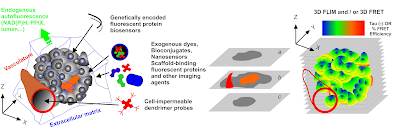
Together with Prof. Margarida Barroso (Albany Medical College, NY) and Prof. Xavier Intes (Rensselaer Polytechnic Institute, NY) we wrote a review article summarising biological aspects of applications of the fluorescence (FLIM) and phosphorescence (PLIM) lifetime imaging microscopies.  We covered most of FLIM and PLIM applications, including FLIM-FRET, macro-FLI, O2 and metabolic imaging, monitoring virus entry into the cell, imaging in organoids and tissue engineering and other areas. In addition, we outlined broad applicability of this type of microscopies not only for cancer and stem research (biomedicine) but also for fungal, plant and other 'general biology' areas.  The review published in the Journal of Cell Science can be assessed here . Twitter link  and link2