Analysis of cell cycle (S phase) without the need of fixation and antibodies
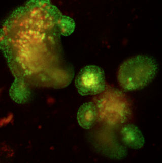
Dr. I. Okkelman, Dr. R. Dmitriev, Prof. D. Papkovsky and T. Foley (Dept. of Anatomy and Neuroscience) have published article describing new method for analysis of cell cycle in live cells. It is based on the discovery that fluorescence lifetime of conventional DNA-staining dye Hoechst 33342 changes upon incorporation of 5-bromo-2-deoxyuridine (BrdU). This allows simple, direct and real-time analysis of live cells, growing in broad range of tissue cultures, from conventional adherent to complex 3D organoid models. The method must be also compatible with in vivo imaging, two-photon excitation and other state-of-the-art imaging platforms and must represent important framework for applications of live cell FLIM in life science areas. It should also inspire other research groups on design of better and more compatible with live cell imaging DNA-binding fluorescent dyes. The team has demonstrated applicability of the method in study of the effect of anti-cancer and life-prolonging drug metformin on the culture of mouse intestinal organoids.
The research has been published in high impact PLoS One journal (ranked number 25 across all disciplines, by Google Scholar Metrics in 2016).
The free full text is available here.


Comments
Post a Comment