Posts

Showing posts from September, 2019

A new preprint from the lab!

Image
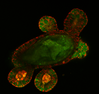
Our team has completed research project focusing on comparison of different methodologies to assess cell bionergetics in the intestinal organoid culture. In order to do that, we analysed Lgr5-GFP mouse organoids (courtesy of Prof. H. Clevers group, Hubrecht Institute) with previously established O2-PLIM method, microplate reader-based Agilent XF96 assay and two-photon excited NAD(P)H-FLIM, in collaboration with Prof. M. Monaghan's group (Trinity College Dublin). For the first time, we found that oxygenation heterogeneity of organoids has functional meaning and is dependent on the metabolic function of stem cell niche. We also observed that stem cells grown in organoids can quickly respond to changes in glucose content in the growth medium. The current preprint version of the manuscript can be found on BioRxiv ( picture above: NADH-FLIM image of live Lgr5-GFP organoid produced on two-photon FLIM microscope Leica Dive SP8 Falcon. Different colors reflect fluorescence lif

SFI Industry Fellowship award for Dr. Ruslan Dmitriev

Image
Dr. Ruslan Dmitriev has received award from the Science Foundation Ireland supporting his industrial fellowship. This fellowship will support continuation of the previous research project on cellulose biosensor scaffold materials within an industrial setting. This is third grant award from the Science Foundation Ireland for Dr. Dmitriev: in the past he also secured SFI Technical Innovation Development Award (TIDA) and Starting Investigator Research Grant (SIRG). More on cellulose biosensor scaffold materials can be found here . Stay tuned for further news!

Estimation of the Mitochondrial Membrane Potential Using Fluorescence Lifetime Imaging Microscopy

Image
Our team has published a new study entitled 'Estimation of the mitochondrial membrane potential using fluorescence lifetime imaging microscopy' in Cytometry Part A journal. In this manuscript we describe the use of well-known green fluorescent SYTO and TMRM dyes in FLIM microscopy: although TMRM has been extensively used in intensity-based readout for the analysis of mitochondrial membrane potential, it has been rarely applied in a fluorescence lifetime domain. We found that reported SYTO 16, 24, TMRM and related dyes enable semi-quantitative assessment of mitochondrial membrane potential in 3D tissue model of mouse intestinal organoids. The paper also shows how the differences in mitochondrial membrane polarization can be visualised within the stem cell niche and at the single cell level using FLIM. The manuscript can be assessed here . Twitter link .