Live cell imaging of mouse intestinal organoids reveals heterogeneity in their oxygenation
 |
| Okkelman et al, Biomaterials, 2017 |
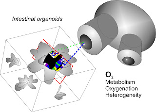
Stem cells extracted from small intestine can grow in the culture dish, to produce mini-organ model termed organoids. They are promising for better understanding stem cell biology, developing drug therapies and success of regenerative medicine. The team led by Science Foundation Ireland (SFI)-funded Starting Investigator Dr. Ruslan Dmitriev studied the dynamics of oxygen supply in live intestinal organoids using method of phosphorescence lifetime imaging microscopy (PLIM) and previously developed O2-sensitive small molecule conjugate. Dr. I. Okkelman, T. Foley, Prof. D. Papkovsky and Dr. R. Dmitriev found that organoids can display dramatic differences in their oxygenation and dynamic gradients, reflecting the heterogeneity of their metabolism and cellular composition. The work published in Biomaterials journal points at the importance of live microscopy analysis of engineered organoid tissue in overcoming the issues of heterogeneity, variability and data reproducibility.
The article text can be accessed here.
The free download link (expires on 31-Oct-2017): https://authors.elsevier.com/a/1ViXTWWN0almS


Comments
Post a Comment