Microscopy image by lab member in the commercial calendar
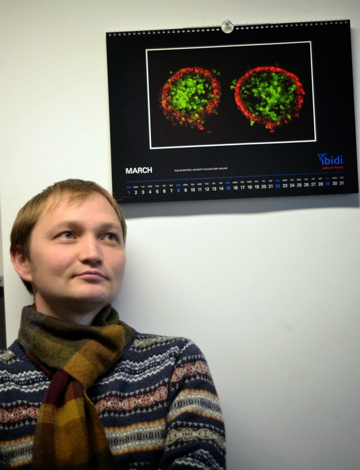
The fluorescence microscopy image of two neurospheres taken by Dr. Ruslan Dmitriev was chosen to appear on 2015 calendar by Ibidi - a well-known company which manufactures consumables for cell and tissue culture. Neurospheres represent free-floating spheroid 3D model of neural stem cells which is characterised by presence of diverse cell types (neural progenitor cells, astrocytes, neurons). They are used for development of better therapies of stroke, studies of inflammation and other neuroscience and stem cell research areas. Dr. Dmitriev and his colleagues from UCC have developed a novel imaging method allowing for real-time analysis of neurosphere oxygenation correlated with cell viability and differentiation. More details on this work can be found here.
This is a second time when microscopy work by Dr. Dmitriev's was chosen for calendars published by Ibidi.


Comments
Post a Comment