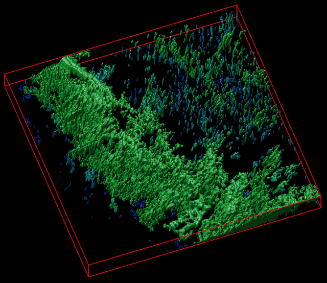
Cellulose-based scaffolds for fluorescence lifetime imaging-assisted tissue engineering
Neil O'Donnell, Dr. Irina Okkelman, Prof. Dmitri Papkovsky and Dr. Ruslan Dmitriev in collaboration with colleagues from the Institute for Regenerative Medicine, Russia have published new research article on the use of various cellulose-based scaffolds for advanced microscopy in tissue engineering applications. The team has developed biosensor based on the recombinant fluorescent biosensing protein and fused with cellulose-binding domain: this cellulose-based pH-sensitive biosensor can label the decellularized plant tissue-derived scaffold (spinach, celery) and is useful for monitoring of extracellular acidification of the 3D culture of cancer cells and stem cell-derived intestinal organoids. Such biomaterial is also compatible with multiparametric FLIM-PLIM imaging of real-time oxygenation providing new framework for the label-free biomedical imaging of the tissue engineered in vitro and ex vivo.
The research published in Acta Biomaterialia journal can be assessed here.
Twitter LinkedIn
Article in the news (Phys.org)
Free download (50 days only) for the final manuscript


Comments
Post a Comment