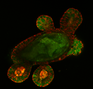
Estimation of the Mitochondrial Membrane Potential Using Fluorescence Lifetime Imaging Microscopy
Our team has published a new study entitled 'Estimation of the mitochondrial membrane potential using fluorescence lifetime imaging microscopy' in Cytometry Part A journal. In this manuscript we describe the use of well-known green fluorescent SYTO and TMRM dyes in FLIM microscopy: although TMRM has been extensively used in intensity-based readout for the analysis of mitochondrial membrane potential, it has been rarely applied in a fluorescence lifetime domain. We found that reported SYTO 16, 24, TMRM and related dyes enable semi-quantitative assessment of mitochondrial membrane potential in 3D tissue model of mouse intestinal organoids. The paper also shows how the differences in mitochondrial membrane polarization can be visualised within the stem cell niche and at the single cell level using FLIM.
The manuscript can be assessed here.
Twitter link.


Comments
Post a Comment