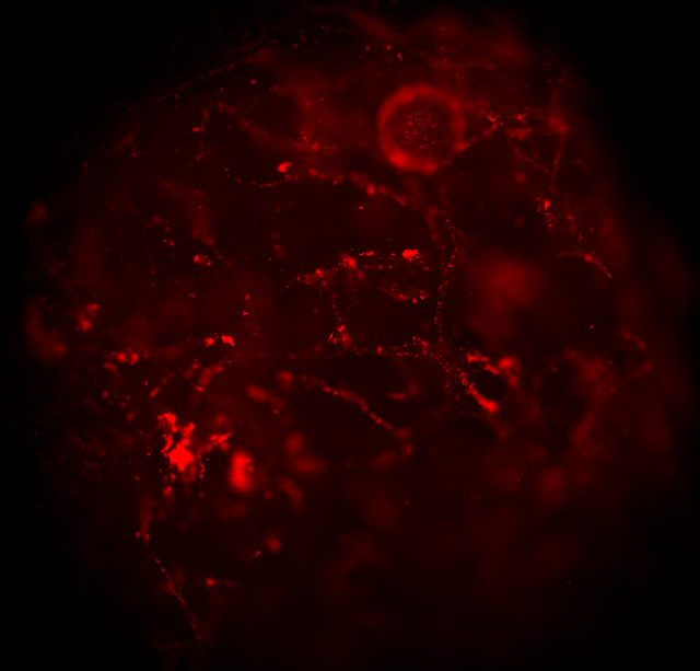
New publication from the group: imaging of O2 nanosensors in implanted scaffold materials
Dr. R. Dmitriev performed and co-authored a collaborative study together with groups from Russia (Sechenov Institute for Regenerative Medicine and Privolzhsky Research Medical University) and Austria (TU Graz), which describes multi-parameter imaging of decellularized pericardial biomeshes in an implanted mouse model. One of highlights of the study is the finding that conjugated polymer-based O2 nanosensors can bind and remain bound to the biomeshes in live animal for extended period of time and thus used for subsequent O2 imaging in live animal and ex vivo. This confirms high usability of conjugated nanoparticle sensors previously developed by our team for such emerging applications as monitoring of tissue regeneration, tumour imaging and others.


Comments
Post a Comment